Positron Emission Tomography (PET) scanning represents one of the most advanced diagnostic imaging technologies available in modern medicine. This comprehensive guide explores everything you need to know about PET scans, from their fundamental principles to their revolutionary applications in detecting disease at the cellular level.
What is a PET Scan?
PET is a common imaging technique, a medical scintillography technique used in nuclear medicine. A radiopharmaceutical—a radioisotope attached to a drug—is injected into the body as a tracer. Unlike traditional imaging methods that primarily show anatomical structures, PET scans reveal functional and metabolic activity within the body’s tissues and organs.
PET is a type of nuclear medicine procedure that measures metabolic activity of the cells of body tissues. Used mostly in patients with brain or heart conditions and cancer, PET helps to visualize the biochemical changes taking place in the body.
According to recent market research, the total volume of PET scans increased 10.2% year over year in 2023, demonstrating the growing reliance on this advanced imaging technology in healthcare settings worldwide.
How PET Imaging Works: The Science Behind the Technology
The Physics of PET Scanning
When the radiopharmaceutical undergoes beta plus decay, a positron is emitted, and when the positron interacts with an ordinary electron, the two particles annihilate and two gamma rays are emitted in opposite directions. These gamma rays are detected by two gamma cameras to form a three-dimensional image.
This sophisticated process allows medical professionals to visualize metabolic processes in real-time, providing unprecedented insight into how organs and tissues function at the cellular level.
Radiopharmaceuticals and Tracers
Radiotracers can be injected, swallowed, or inhaled depending upon the site of the body being examined, and the tracer gets absorbed by various tissues according to their specific affinity. The most commonly used radiotracer is fluorodeoxyglucose (FDG), which mimics glucose and accumulates in areas with high metabolic activity.
Research indicates that 82% of PET scans used F-18 FDG, 7% used rubidium-82, and all other agents made up the remainder of radiopharmaceutical usage in recent studies.
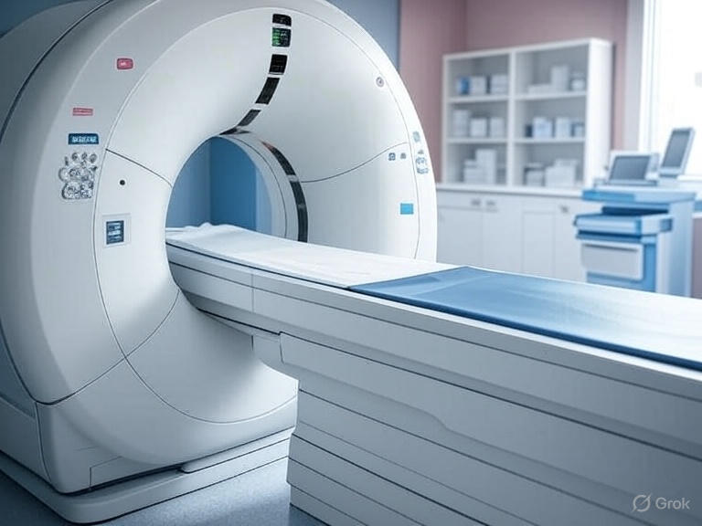
Advanced PET Technology: PET-CT and Hybrid Imaging
The Evolution to PET-CT Systems
PET scanners can incorporate a computed tomography scanner (CT) and are known as PET–CT scanners. This combination provides both functional and anatomical information in a single examination, significantly improving diagnostic accuracy.
Most PET scans are now performed along with a CT scan. This combination scan is called a PET/CT. This helps find the exact location of the tumor or other abnormality.
Digital PET Innovation
In the future, 2025 to 2035, the tendency will be towards digital PET scanners offering images of better quality with reduced scanning time and, moreover, the growing need for PET as part of personalized medicine and theranostics, particularly in novel oncology drug development, represents a significant advancement in medical imaging technology.
The improvements include new hardware, incorporating designs with digital photomultipliers, and fast electronics, allowing implementation of time-of-flight reconstruction. Manufacturers also improved PET sensitivity with a larger axial field of view and 3D imaging.
Clinical Applications of PET Scanning
Oncology: Cancer Detection and Monitoring
PET scans have revolutionized cancer care by enabling early detection and precise monitoring of treatment response. Unlike other imaging techniques, PET scans focus on processes and molecular activity within your body. This gives them the potential to identify cancerous cells before structural changes become apparent on conventional imaging.
By identifying changes at the cellular level, PET may detect the early onset of disease before other imaging tests can. This early detection capability is particularly valuable in oncology, where timely intervention can significantly impact patient outcomes.
Cardiac Applications
Identify areas of the heart muscle that would benefit from angioplasty or coronary artery bypass surgery. PET scans provide detailed information about cardiac metabolism and blood flow, helping cardiologists make informed treatment decisions.
It can help detect heart disease and other conditions. The scan typically takes 30–60 minutes.
Neurological Imaging
If you’re experiencing neurological symptoms, your provider may recommend a PET scan to evaluate possible brain abnormalities, such as tumors, seizures and other central nervous system conditions.
PET neuroimaging has proven particularly valuable in diagnosing and monitoring conditions such as Alzheimer’s disease, Parkinson’s disease, and epilepsy, where metabolic changes often precede structural abnormalities.
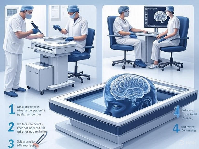
PET Scan Procedure: What Patients Can Expect
Pre-Scan Preparation
Tell your doctor if there is any possibility you are pregnant or you are breastfeeding. Your doctor will tell you how to prepare based on the type of your exam. Discuss any recent illnesses, medical conditions, medications you are taking and allergies – especially to contrast material.
For FDG PET scans, patients typically need to fast for 4-6 hours before the procedure to ensure optimal tracer uptake. Blood sugar or insulin levels may affect the test results in people with diabetes.
During the Scan
The PET scanning process involves several key steps:
- Tracer Administration: The radiotracer is administered intravenously, orally, or through inhalation
- Uptake Period: Patients wait 30-90 minutes for the tracer to distribute throughout the body
- Image Acquisition: The actual scanning process takes 20-40 minutes
- Post-Processing: Advanced computer algorithms reconstruct detailed 3D images
PET scans help a doctor diagnose certain health conditions, plan treatment, find out how an existing condition is developing, or check the effectiveness of a treatment.
Benefits and Advantages of PET Imaging
Early Disease Detection
The primary advantage of PET scanning lies in its ability to detect diseases at the molecular level before anatomical changes occur. This capability is particularly crucial in cancer diagnosis, where early detection significantly improves treatment outcomes.
Functional vs. Structural Imaging
While traditional imaging modalities like CT and MRI excel at showing anatomical structures, PET scans provide unique insights into tissue function and metabolism. This functional information helps clinicians understand not just where a problem exists, but how it affects normal physiological processes.
Treatment Monitoring
PET scans enable precise monitoring of treatment response by showing changes in metabolic activity. This capability allows healthcare providers to adjust treatment plans quickly based on objective evidence of therapeutic effectiveness.
Limitations and Considerations
Radiation Exposure
As a nuclear medicine procedure, PET scanning involves exposure to ionizing radiation. However, the radiation doses are carefully calculated to be as low as reasonably achievable while maintaining diagnostic quality.
Cost Considerations
PET scans are more expensive than conventional imaging studies due to the sophisticated technology and short-lived radiopharmaceuticals required. The Positron Emission Tomography Market size is expected to reach USD 1.13 billion in 2025 and grow at a CAGR of 3.71% to reach USD 1.36 billion by 2030, reflecting both the technology’s value and its associated costs.
Tracer Availability
The short half-life of many PET tracers requires specialized production facilities and careful scheduling to ensure optimal imaging quality.
Market Growth and Future Trends
Current Market Dynamics
Looking ahead, 47% of PET imaging sites anticipate their scan volumes will increase over the next 12 months, 44% of volumes to stay the same, and 1% anticipate a decrease. This growth reflects increasing recognition of PET’s clinical value across multiple medical specialties.
Expanding Accessibility
Adoption in Diagnostic Centers: Lower costs and enhanced accessibility are driving diagnostic centers to integrate PET scanners into their services. This trend increases local access to advanced imaging for patients.
Technological Advancements
Future developments in PET technology focus on improving image quality while reducing scan times and radiation exposure. Digital PET systems and artificial intelligence integration promise to further enhance diagnostic capabilities.
PET vs. Other Imaging Modalities
PET vs. MRI
While MRI provides excellent soft tissue contrast and anatomical detail, PET offers unique metabolic information. PET-MRI hybrid systems combine these complementary strengths for comprehensive diagnostic evaluation.
PET vs. SPECT
Both PET and SPECT are nuclear medicine imaging techniques, but PET offers superior spatial resolution and sensitivity. PET’s ability to provide quantitative measurements makes it particularly valuable for research and treatment monitoring.
PET vs. CT
CT scans excel at showing anatomical structures and bone abnormalities, while PET reveals functional information. The combination of PET-CT provides both anatomical and functional data in a single examination.
Safety Considerations and Patient Care
Radiation Safety
Modern PET scanners incorporate advanced safety features and dose optimization protocols to minimize radiation exposure while maintaining image quality. Regulatory bodies continuously monitor and update safety guidelines for nuclear medicine procedures.
Special Populations
Pregnant women and breastfeeding mothers require special consideration before PET scanning. Alternative imaging methods may be preferred unless the PET scan is essential for diagnosis or treatment planning.
Pediatric Considerations
Children may require specialized protocols including sedation for some procedures. Weight-based dosing ensures appropriate tracer administration while minimizing radiation exposure.
Interpreting PET Scan Results
Quantitative Analysis
PET scans provide quantitative data through standardized uptake values (SUVs) and other metrics that allow objective assessment of metabolic activity. These measurements help clinicians make evidence-based treatment decisions.
Multidisciplinary Review
Complex PET scans often require multidisciplinary review involving nuclear medicine physicians, radiologists, oncologists, and other specialists to ensure comprehensive interpretation and optimal patient care.
Future Directions in PET Imaging
Artificial Intelligence Integration
Machine learning algorithms are increasingly being integrated into PET imaging workflows to improve image reconstruction, reduce noise, and assist with interpretation. These developments promise to enhance both efficiency and diagnostic accuracy.
Novel Radiopharmaceuticals
Research continues to develop new PET tracers targeting specific biological processes and disease pathways. These specialized tracers may enable more precise diagnosis and treatment monitoring for various conditions.
Personalized Medicine
the growing need for PET as part of personalized medicine and theranostics, particularly in novel oncology drug development represents an exciting frontier where imaging directly guides therapeutic decision-making.
Conclusion
PET scanning has established itself as an indispensable tool in modern medicine, offering unique insights into cellular function and metabolism that complement traditional anatomical imaging. As technology continues to advance, PET imaging will likely play an even more significant role in early disease detection, treatment planning, and personalized medicine.
The continued growth in PET scan volumes, technological improvements, and expanding clinical applications demonstrate this imaging modality’s essential role in contemporary healthcare. For patients and healthcare providers alike, understanding PET scanning capabilities and limitations enables optimal utilization of this powerful diagnostic tool.
Whether used for cancer detection, cardiac assessment, or neurological evaluation, PET scans provide critical information that helps guide treatment decisions and improve patient outcomes. As we move forward, the integration of artificial intelligence, development of novel tracers, and improvements in scanner technology promise to further enhance the diagnostic power of PET imaging.
For healthcare professionals seeking to optimize patient care and for patients facing diagnostic challenges, PET scanning represents one of medicine’s most sophisticated approaches to understanding disease at the molecular level. This technology continues to push the boundaries of what’s possible in medical diagnosis, offering hope for earlier detection and more effective treatment of various conditions.
For more information about advanced medical technologies and their applications, you might find our comprehensive guide on German Shepherds interesting for pet-related health monitoring technologies. Additionally, explore innovative diagnostic tools at Snapspeak for cutting-edge healthcare solutions.
References: Information in this article is sourced from peer-reviewed medical literature, authoritative medical institutions including Mayo Clinic, Cleveland Clinic, Johns Hopkins Medicine, and medical databases such as PubMed and Wikipedia. Market data is derived from industry reports and professional medical imaging organizations.